Glutaric Aciduria Type-1is a type of inherited metabolic disorder due to deficiency of glutaryl-CoA-dehydrogenase. The glutaryl-CoA-dehydrogenase is responsible for breakdown of L-lysine, L-hydroxylysine & L-tryptophan leading to elevated glutaric acid, 3 hydroxyl glutaric acid, glutaconic acid & glutaryl carnitine1.
New born with this condition may born healthy or with nonspecific findings like macrocephaly & hypotonia. Acute metabolic crisis may develop following: Febrile illness, gastroenteritis or immunization1. The crisis may trigger stiatal injury producing distonia, dyskinesia, chorioathetosis, cognitive impairment & coma. Patient may have intracranial haemorrhage, speech problem & seizure.
Magnetic resonance imaging of the brain is the modality of choice to investigate children with possible GA-1. MRI shows bilateral symmetrical basal ganglia T2 & FLAIR hyperintensity which appear hypointense on T1 weighted images. T2 & FLAIR hyperintense lesions are also noted in substantia nigra, dentate nucleus. Symmetrical T2 white matter hyperintensities are also observed in periventricular & subcortical regions. The lesions in the acute stage show diffusion restriction with progressive atrophy in the chronic stage. No demonstrable susceptibility change or contrast enhancement is seen in the lesions. MR spectroscopy shows presence of lactate peak in acute lesions. Common presentations include enlarged extraaxial CSF spaces predominantly in the anterior temporal regions, wide sylvian fissures & ventricular enlargement. Wide sylvian fissures reveal typical Batwing appearance2. There may be subdural haematoma possibly due to stretching of bridging veins in the enlarged extraaxial fluid space3. Enlargement of optic chiasm with signal alteration in the anterior intracranial visual pathway has also been reported rarely4.
Diagnosis is confirmed by elevated excretion of glutaric acid, three hydroxyl glutaric acid & hypocarnitaemia.
Differential diagnosis includes conditions having macrocephaly with leukodystrophy like canavan disease, alexander disease & megalencephalic leukoencephalopathy with cyst. Symmetrical T2 hyperintense basal ganglia lesions like some aminoacidopathies, mitochondrial disorders & Wilson disease may also mimic Glutaric Aciduria Type-15. Benign external hydrocephalus is another condition which needs to be differentiated.
Treatment: Restriction of gluterigenic aminoacid, lysine, tryptophan & hydroxylysine. There should be supplementation of carnitine & riboflavin.
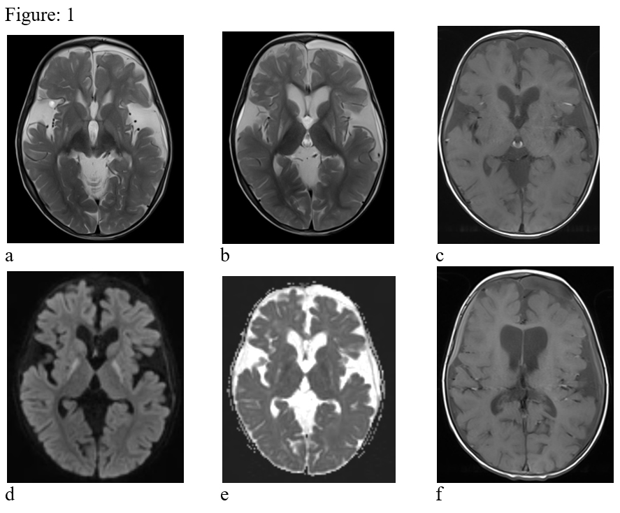
Figure:1- T2 hyperintense & T1 hypointense symmetrical lesions in bilateral basal ganglia regions & widening of the sylvian fissures bilaterally (a, b & c). The basal ganglia lesions reveal restricted diffusion on DW images (d & e). Subdural haematoma (acute to chronic) is seen in bilateral fronto-temporal & left parietal regions (f).
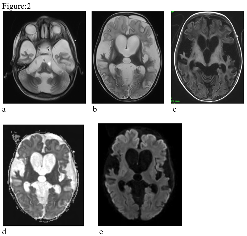
Figure:2- Follow-up MRI after 1 year. There is progressive atrophy of the supra & infratemporal brain parenchyma (a, b & c). The diffusion restriction is not seen in present study (d & e).
References:
- Nunes, S. Loureiro, S. Carvalho, R.P. Pais, C. Alfaiate, A. Faria, P. Garcia, & L. Diogo. Brain MRI Findings as an Important Diagnostic Clue in Glutaric Aciduria Type-1. Neuroradiol J.2013 Apr; 26(2): 155–161.
- Anusha Doraiswamy, Bhanu Kesavamurthy, Lakshminarasimhan Ranganathan. Batwing appearance – A neuroradiologic clue to glutaric aciduria-type-1. International Journal of Epilepsy, Volume 2, Issue 1, January–June 2015, Pages 44-48
- Gary L. Hedlund, Nicola Longo, Marzia Pasquali. Glutaric Acidemia Type-1. Am J Med Genet C Semin Med Genet. 2006 May 15; 142C(2): 86–94
- A. Ntorkou, J. Daire, F. Renaldo, D. Doummar, M. Alison, M. Schiff and M. Elmaleh-Bergès. Enlargement of the Optic Chiasm: A Novel Imaging Finding in Glutaric Aciduria Type-1, American Journal of Neuroradiology, 2021 September, 42 (9) 1722-1726.
- Hajar Zebbakh, Ibrahima Diallo, Najlae Lrhorfi, Dina Alami, Nazik Allali, Latifa Chat. Glutaric aciduria type 1: Typical aspects in imaging. Edorium J Radiol 2022; 8(2):5–9.