t consequat semper viverra nam libero. Lorem dolor sed viverra ipsum nunc aliquet bibendum.
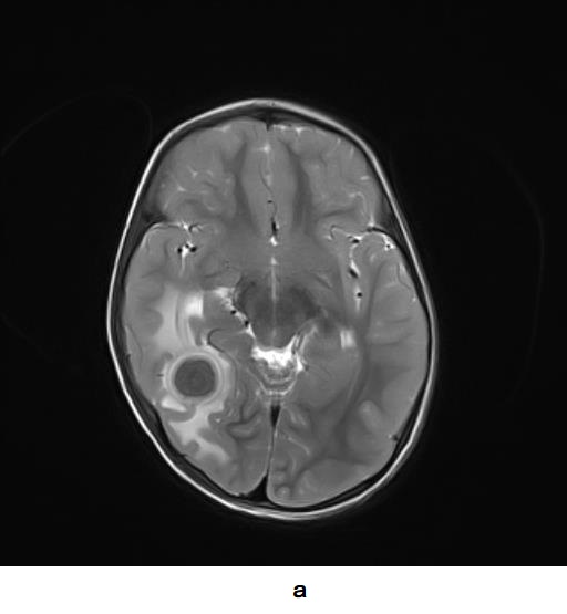
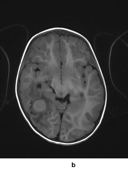
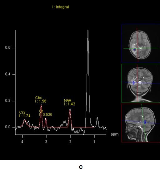
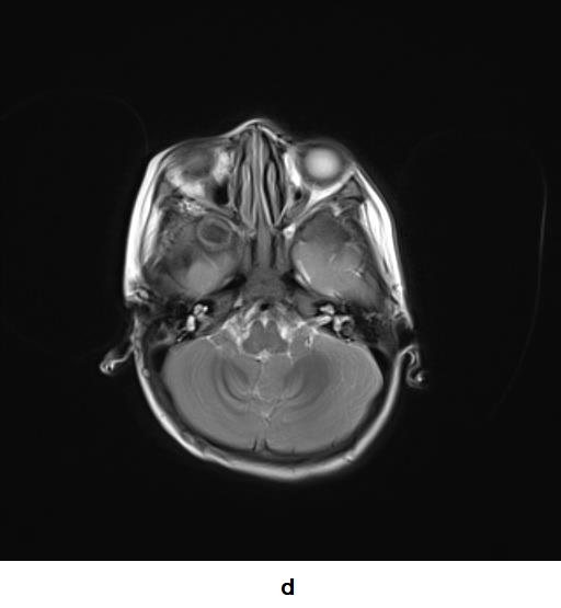
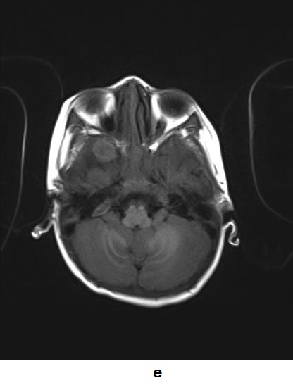
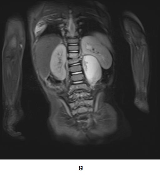
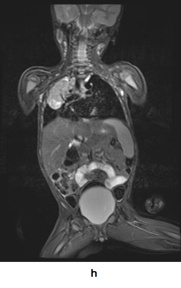
2 yrs male patient presenting with features of raised ICP. There is H/O fever & chronic cough. There is T2 hypointense & T1 iso to hyperintense well defined lesion with hyperintense rim on T2 & perifocal edema (a, b). MR spectroscopy shows large lipid peak at 1.3 ppm (c). Well defined T2 hypointense lesion is seen in right sphenoid wing (d) which is mildly hyperintense on T1 weighted images (e). There is evidence of Koch’s spine (f, g) and pulmonary lesion on the right side (h).